Prof. Suhono Harso Supangkat
STE-ITB
Gitarja Sandi
STEI-ITB
Arga Daniel Reynardo Samosir
STEI-ITB
Alya Apriliyanti
STEI-ITB
Abstract
Teleradiology, as part of this advancement, allows radiological images such as X-rays, MRIs, and CT scans to be transmitted and analyzed remotely. This is very beneficial in personalized healthcare as it speeds up the process of diagnosis and treatment. With teleradiology, radiologists can provide fast and accurate interpretations and recommendations, regardless of the physical location of the patient or the doctor. This is especially helpful in cases of medical emergencies and for patients in remote areas.
Medical image analysis tasks are performed repeatedly, leading to fatigue and reduced capabilities of radiologists. This research applies the DSRM by developing a solution design to address the challenges faced by doctors in medical image analysis and testing this design against the needs of radiologists. The solution developed is an automation prototype to generate AI analysis reports in DICOM Structured Report (DICOM-SR) format on DICOM storage, specifically for chest X-ray (CXR) images. This prototype successfully automates the generation of AI analysis reports in DICOM Structured Report (DICOM-SR) format.
Keyword: Teleradiology, DICOM, LLM
Introduction
In the modern medical era, personalized healthcare has become a major focus in research and clinical practice. This development is driven by the need to address genetic diversity and individual responses to treatment, especially amidst the increasing prevalence of chronic diseases. Personalized healthcare aims to provide treatment tailored to the specific needs of each patient, based on their genetic profile, environment, and lifestyle.
Teleradiology has brought a new dimension to personalized healthcare. Its ability to remotely transmit radiological images such as X-rays, MRIs, and CT scans has accelerated the process of diagnosis and treatment, especially for patients in remote areas. However, this also poses challenges such as the need for robust infrastructure, reliable connectivity, and questions about the accuracy and reliability of image interpretation by AI and machine learning.
Research Objectives
● Designing and building a prototype teleradiology system that can support personalized healthcare in telemedicine services.
● Analyzing teleradiology so that it can improve the accuracy and speed of diagnosis in health services that support personalized healthcare.
● Designing the integration of radiology data with clinical information in the EHR to support a more personalized treatment approach.


Discussion & Result
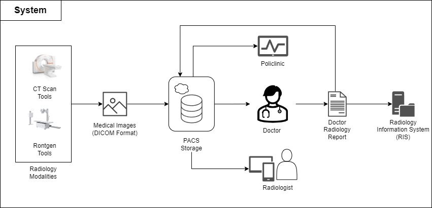
The Medical Image Analysis activity is performed repeatedly, leading to several problem points: The doctor’s ability to focus decreases as the number of cases handled increases. The accumulation of cases creates waiting times, preventing patients from receiving prompt treatment.
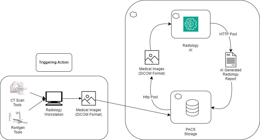
AI analysis can assist the radiology field by providing a second opinion on diseases, information obtained more quickly, and information provided in a complete and comprehensive manner
Proposed Solution

Conclusion
The prototype of the automation of AI analysis report generation on DICOM storage was successfully implemented. The implementation was carried out by combining and integrating the work of Orthanc and AI with the LLaVA-Med model. All functional requirements that had been set were successfully achieved by the system. This result indicates that the needs of radiologists can be met, and by developing and implementing this prototype, radiologists can be helped to perform analysis. The benefits obtained by helping doctors are that the analysis results can be obtained quickly and accurately so that patient care can be carried out faster and more precisely.
This research is supported by PPMI Program and Smart City & Community Innovation Center